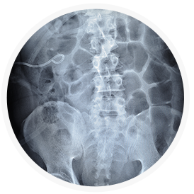
Ureteroscopy
Ureteroscopy is the technique of choice with smaller stones that lodge in the mid and lower sections of the ureter. A ureteroscope (flexible, fiberoptic instrument resembling a long, thin telescope) is inserted through the urethra and bladder to the stone. The urologist can locate the stone visually and remove it with a small basket-like device inserted through the ureteroscope. This procedure is called endoscopic basket extraction. A laser may be used to break the stone into smaller pieces, which can be passed by the patient. Ureteroscopy is performed under general or local anesthesia on an outpatient basis. The urologist often places a small silicone tube (stent) into the ureter to relieve swelling and facilitate healing.
Lithotripsy
A variety of nonsurgical techniques have been developed to crush or pulverize kidney stones. Lithotripsy uses a machine called a lithotripter to project shock waves or sonic pulses against the stone and break it into tiny particles that can pass naturally in the patient’s urine. This can be done in several ways, depending on the size and location of the stone.
Patients undergoing lithotripsy are given a sedative and a general or local anesthetic. Shock waves are focused on the kidney stone at a rate of approximately one per second and the therapy may last over an hour. Bruising may result from the shock waves and discomfort may be experienced as the crushed calculi are passed; but, most patients resume normal activity in a few days.
Lithotripsy is highly effective for stones in the kidney and upper ureter. More than one treatment may be required. Rarely, a catheter may be inserted through a small incision in the back to drain the kidney and remove the stone fragments. Patients with very large stones or complicating medical conditions may require different treatment.
Percutaneous Stone Removal
Also known as (Percutaneous Nephrolithotomy, or PCNL). Some kidney stones cannot be treated by ESWL or Ureteroscopy (see above) for a variety of reasons. Stones that are large (>2cm), impacted stones of the kidney or ureter, or “staghorn” stones of the kidney often cannot be treated with less invasive procedures. A PCNL involves two steps – usually done the same day. The first is the placement of a nephrostomy tube under light anesthesia by the intervention radiologist in the radiology suite. A nephrostomy tube is a thin (3mm) tube that enters the kidney directly through the skin in the back or flank. The second step is the actual breaking and removal of the stones (nephrolithotomy) under general anesthesia by the urological surgeon in the operating room. The PCNL has a very high success rate of removing all the stones in one procedure. Post-operatively, a nephrostomy tube is left in place (in the kidney) in addition to a Foley catheter (in the bladder). These are usually removed within the first 48 hours and the patient is usually discharged from the hospital in 1-2 days.


